Fig. 2

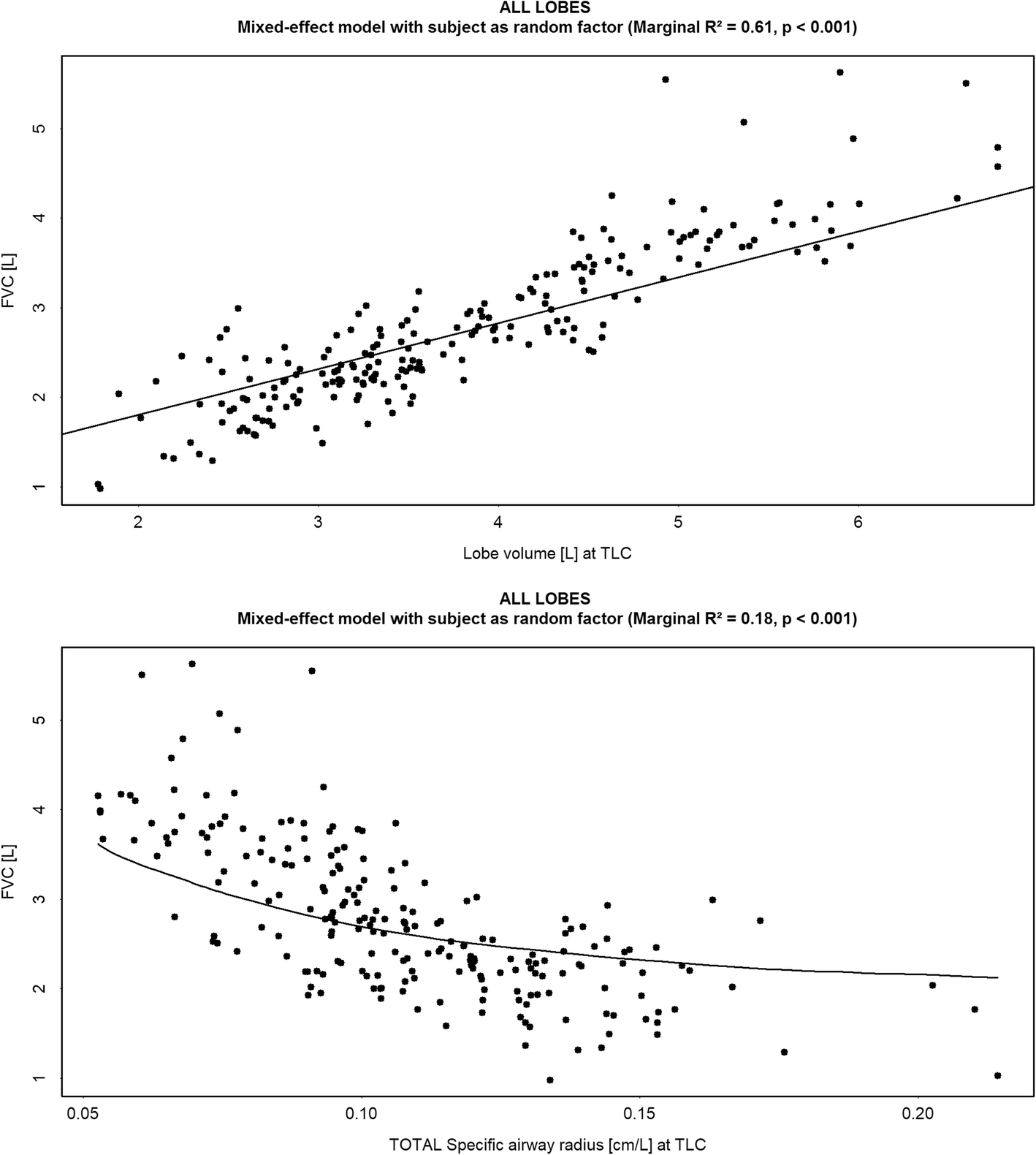
(Upper panel) Correlation between the FRI-based lung volume measured at Total Lung Capacity (TLC) [L] and the Forced Vital Capacity (FVC) [L]; (Lower panel) Correlation between the specific image based airway radius (siRADaw) measured at Total Lung Capacity (TLC) [cm/L] and the Forced Vital Capacity (FVC) [L]