Fig. 2
From: Ciliated conical epithelial cell protrusions point towards a diagnosis of primary ciliary dyskinesia

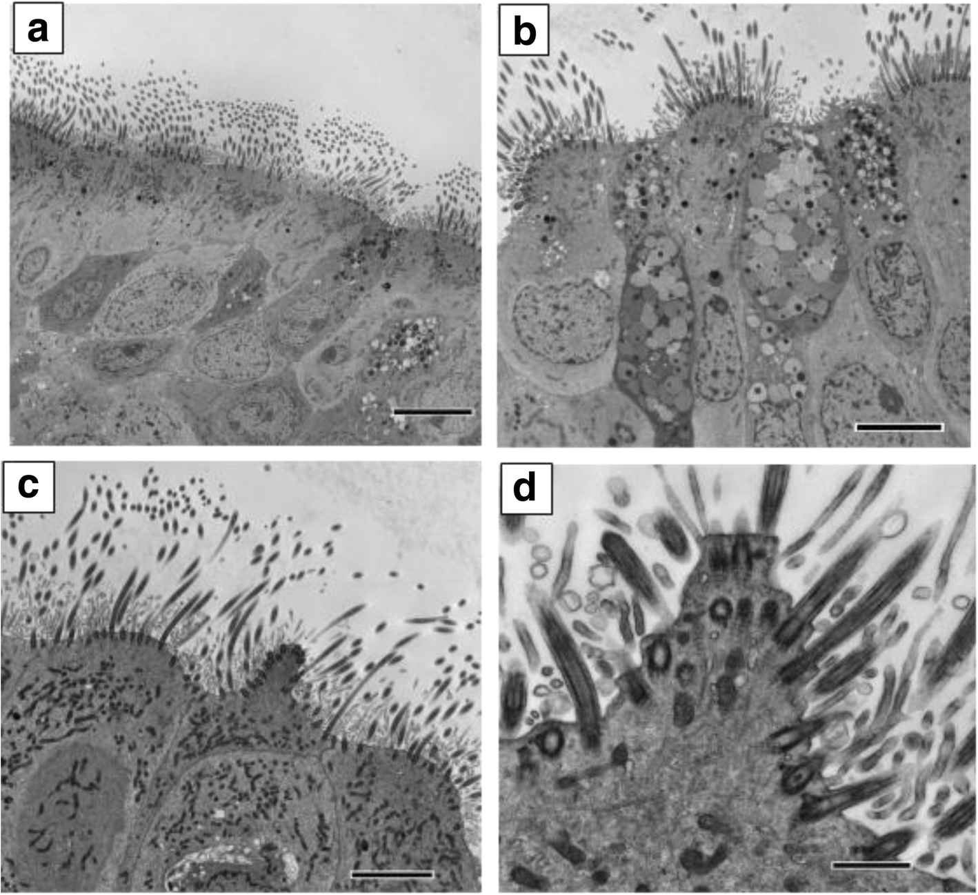
Transmission electron microscopy cross sections of ciliated respiratory epithelium showing; a Healthy well ciliated epithelium from a healthy control (bar = 8 μm). No protrusions were seen in the TEM images obtained from CF and Asthma patients. b Ciliated respiratory epithelium from a patient with bronchiectasis who had the diagnosis of PCD excluded. Ciliated cells are seen to project from the epithelium. Note the projection involves the whole surface of the cell and does not have a conical shape (bar = 6 μm). c Ciliated epithelium from a patient with PCD showing a characteristic ciliated conical protrusion (arrow) (bar = 4 μm). d High power of a ciliated conical protrusion (bar = 800 nm)